Glaucoma - Progression
How can OCT help us track the progression of glaucoma?
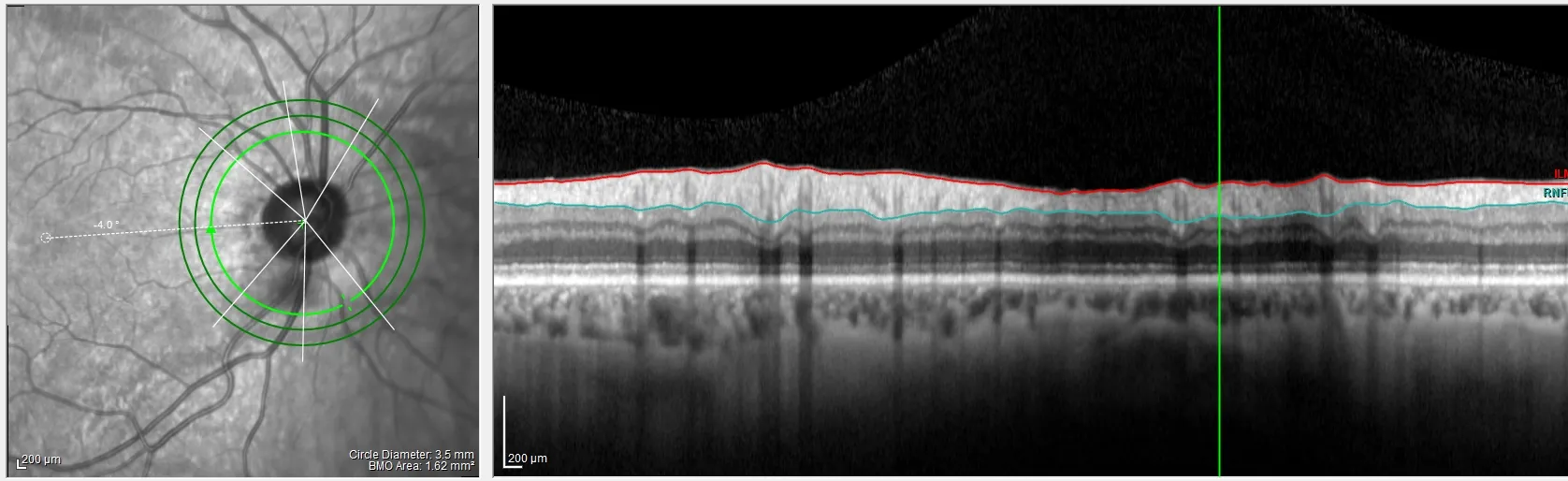
One of our research focuses is to understand and help develop techniques for tracking the progression of glaucoma in clinical practice. We create models, as well as use qualitative methods, in order to understand the best methods for tracking structural and functional changes in vision and visual anatomy.
Currently the main goal of our work is to better understand the nature of glaucomatous damage and its progression, as well to improve the ability of the clinician to quickly and accurately detect glaucomatous damage, and its progression in a clinical setting. Key to this work is a comparison of the behavioral changes, as measured with visual fields and automated perimetry, and anatomical changes as measured with optical coherence tomography (OCT) associated with glaucoma. Our goal here is nothing short of changing the way clinicians use OCT and visual fields to detect and follow glaucomatous damage in the clinic. One OCT company has already incorporated a “Hood Report” into their software and another is about to do the same.
Relevant Recent Reviews/Papers:
- Hood DC, De Moraes CG. (2018) Challenges to the Common Clinical Paradigm for Diagnosis of Glaucomatous Damage With OCT and Visual Fields. Invest Ophthalmol Vis Sci. 2018;59(2):788-791.
- Hood, DC. (2017) Improving our understanding, and detection, of glaucomatous damage: An approach based upon optical coherence tomography (OCT). Progress in Retinal and Eye Research. 57:46-75.
- Hood DC, De Cuir, N, Blumberg DM, Liebmann J, Ravivarn Jarukasetphon, Ritch R., De Moraes CG, (2016) A single wide-field OCT protocol can provide compelling information for the diagnosis of early glaucoma. Translational Visual Science & Technology. 5(60
- Hood DC, Raza AS, De Moraes CG, Alhadeff PA, Idiga J, Blumberg DM, Liebmann JM, Ritch R. (2014) Evaluation of a one-page report to aid in detecting glaucomatous damage. Translational Vision Science & Technology 3, Dec 17;3(6):8.